Anatomy Of Gallbladder Pdf To Jpg
Welcome to PowerPictures - our rapidly expanding line of professional stock photos with over 60 million images to choose from! Whether you are looking for visually-stunning photographs for your next marketing campaign or eye-catching pictures for your website or product brochures, we've got what you need for very low prices.
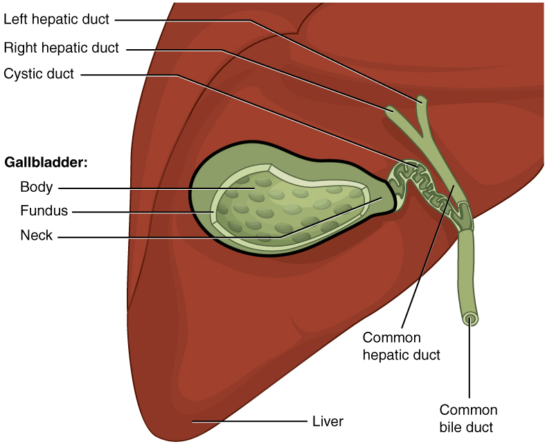
Anatomy and Physiology Gallblader - Download as Word Doc (.doc /.docx), PDF File (.pdf), Text File (.txt) or read online. Gallbladder concentrates bile fivefold. Male to female 1:3 as shown inFigures 6-8. Other pathologies of gallbladder were found to be acute cholecysti-ties 12%, chronic cholocystities 5%, sludge 2%, carcinoma of the Gall-bladder 1% and Gall-bladder polyps 1%. Emphysematous choleycystities 1%.
All images are supplied in the popular JPEG file format and are available in both lower resolutions (suitable for on-screen applications) and various higher resolutions (suitable for high-quality print applications). We also provide a sophisticated search engine to show you the best results for whatever you are searching for.

Not just good photos that happen to use the words you searched on, but actually great photos, sorted to first show the best, most relevant, inspirational, motivational and powerful pictures that other people like you have purchased in the past. And, as you know, that really helps when you're short on time!
Results The urinary bladder wall measured 0.11 ± 0.03 cm. The symmetrical kidneys were in the left and right cranial quadrant of the abdomen and the cortical, medullary and renal pelvis regions were recognized and in all sections. Buku kedokteran gratis pdf lézer nyomtatók. The medullary rim sign was visualized in the left kidney of two coatis. The liver had homogeneous texture and was in the cranial abdomen under the rib cage. The gallbladder, rounded and filled with anechoic content was visualized in all coatis, to the right of the midline. The spleen was identified in the left cranial abdomen following the greater curvature of the stomach. The parenchyma was homogeneous and hyperechogenic compared to the liver and kidney cortex.
The stomach was in the cranial abdomen, limited cranially by the liver and caudo-laterally by the spleen. The left adrenal glands of five coatis were seen in the cranial pole of the left kidney showing hypoechogenic parenchyma without distinction of cortex and medulla.
The pancreas was visualized in only two coatis. The left ovary (0.92 cm x 0.56 cm) was visualized on a single coati in the caudal pole of the kidney. The uterus, right adrenal, right ovary and intestines were not visualized.
The coati ( Nasua nasua) is a member of the Procynidae family of the Carnivorous order [ ]. With the exception of Chile [ ], this is a specimen exclusive of South America. It is an animal that easily adapts and socializes with humans; therefore, most studies related to this species refer to the reproductive area, aiming to control population in reserves and zoos, or with their ecological role of seed dispersal [ ]. As these animals can also be reservoirs for pathogens of diseases such as leishmaniasis, rabies and distemper, there is the concern of transmission of some of these diseases to human populations or domestic animals [ ]. Thus, there is a need for detailed information on aspects related to diseases affecting the coatis and for veterinarians to deepen their knowledge of available diagnostic imaging aids.